From Student to Scientist: A Summer of Hands-On Research
My name is Sophia Serrano, and I am a Mexican/Puerto Rican American from the West side of Chicago interested in pursuing a career in STEM. I am currently five weeks into my Sci-High Summer Program at Northwestern University’s George M. O’Brien Kidney Core Center (NUGoKIDNEY) as a returning SciHigh Scholar. I’ve always had a love and passion for STEM-related fields, especially medical science. This being my second year in the program, I am ecstatic to have the opportunity to return and further my knowledge about scientific research. I have genuinely enjoyed every moment, being able to constantly learn new skills, put my research into practice, and learn about different STEM-related studies and fields throughout the summer.
I was first introduced to the program during my sophomore year when my biology teacher approached me asking if it was something I would be interested in. Knowing that I had an interest and was passionate about pursuing a medical career but not entirely sure where to start, I knew this would be a good opportunity to get my foot in the door. As the program's objective was to contribute and curate a poster presentation that would later be presented in front of a large crowd, I was anxious because of my lack and fear of public speaking. Despite feeling a bit discouraged due to my lack of prior experience in science-related courses and programs, I decided to take the chance and apply. I made sure to do extensive research about the Sci-High program to prepare for what was to come. To no surprise, I was completely intrigued by what the summer ahead of me had in store.
Overview
This summer I have the opportunity to work alongside Benjamin Thomson, PhD, and Elysse Brookins, a member of his Laboratory, trying to identify non-endothelial expression patterns of endomucin. The main point of this study is to learn more about endomucin as a whole and how it can be useful as a molecular marker, as it is a membrane-bound glycoprotein. Different cell types throughout the body can be identified by using different protein expressions. It is important in both research and clinical pathology standpoints to have tools for identifying different cell types accurately. In order for these tools to be useful, we need to know exactly which proteins are where and how we can use them effectively, as different cell types and molecular markers help to differentiate them. These unique patterns of molecular markers and protein expressions are essential to identify different cell types in research as they make sure that they are accurately identifying what they say they are. Endomucin is currently being used as a marker for identifying endothelial cells in different tissues throughout the body. Yet based on our preliminary data sets, it has been proven that endomucin has also been expressed in non-endothelial cells, making the marker not entirely accurate, which can lead to a misinterpretation of results that could affect different studies.
To start our research, we collected different organs from 4 adult mice; this included the kidney, liver, heart, lung, brain, small intestine, eye, pancreas, and spleen. This process in and of itself was very new to me, as this was my first time being in a professional lab setting, and I was not entirely sure what to expect. I watched as the mice were euthanized and each organ was carefully dissected and fixed in a formaldehyde solution, a crucial step in preserving the tissues. They were then placed in paraffin wax to prepare them to be sliced into 5-micron thick sections, allowing us to start staining. We first started with an H&E stain (Hematoxylin and Eosin) just to validate the quality and identify the tissues before we started using the immunofluorescent stains. Immunofluorescent staining is the process of identifying different proteins in the tissue sections by using antibody-based methods to detect different proteins. This helped us identify whether the endomucin was solely expressed in endothelial cells or if they were prominent in different cell types. Doing this entire process was very new to me as this was my first time being in a lab, though with practice and patience I was soon able to complete the protocol without help. This was a process that I enjoyed because I was able to spend most of my time in the lab learning about the process while immersing myself in the environment daily.
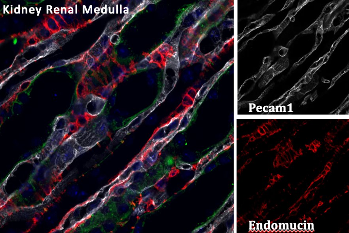
Once the staining was completed and a cover slip was placed on the sample slide to protect the tissues, we were finally able to view them under a microscope. Since we used immunofluorescent staining as our secondary antibody, there is a specific type of microscope that needs to be used to get the highest-resolution images. By using laser scanning confocal microscopy, we were able to specifically see the areas that are expressing endomucin without the confusion of overlapping cells. Due to the different wavelengths of the light that the different antibodies emit, we are given three different images that we can use to vividly show whether or not endomucin is being expressed in non-endothelial cells. The images below show sections of the kidney focused on the renal medulla where you can clearly see the difference between endomucin highlighted in red and Pecam1 (platelet endothelial cell adhesion molecule) highlighted in white.
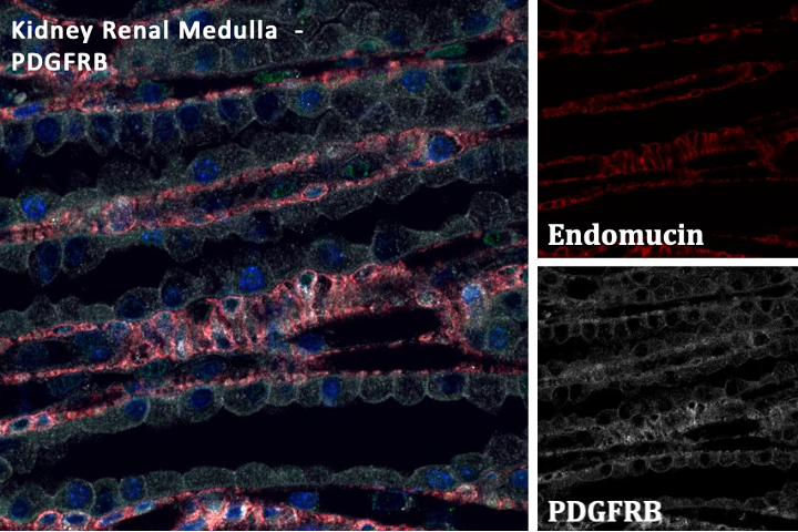
The combination of a high-resolution laser and the pinhole opening that it goes through disperses the laser throughout the tissue giving a high-quality and accurate image. This truly had to be my favorite part of the process by far, as I also have an interest in photography. Being able to combine both my hobby and passion allowed me to use my creative and scientific skills when it came time to process and edit the images.
This being my second year in the program, I used my past knowledge gained during the summer of 2022 to assist. I had the pleasure of working with my mentor Anand Srivastava, MD, MPH, where we investigated the association of Renal Blood Flow, as assessed by CEUS (Contrast Enhanced Ultrasound) with Histopathologic lesions. As Dr. Srivastava conducted a clinical research study, there was zero experience in the lab, therefore most of the knowledge and skills I gained were purely based on Scientific Journals and personal conversations. Due to the lack of hands-on lab experience, I was able to shadow Dr. Srivastava in the clinic, as a patient part of the study was injected with microbubbles to assess its wash-in rate, its overall function, and see if there were any lesions. Since this was my first time learning about kidneys and their functions, I spent most of my time learning how to read and fully understand Scientific journals that related to my poster which were mostly about Chronic Kidney Disease and Glomerular Filtration Rate. Being able to fully immerse myself in my research, I fully understood the study and how it could impact the way we diagnose the severity and longevity of CKD in a non-invasive way. Aside from this, to prepare myself for the end goal of the program, I was instructed to give weekly presentations based on the journals I had read to prove my comprehension and ability to both explain and present. This indefinitely aided me in being able to confidently present during the showcase, as well as giving me countless skills and valuable information I have been able to put into practice this summer.
The Human Side of Science
Going into the program I already had an assumed idea of what the people in the lab were going to be like, knowing that they were very professional individuals that were passionate about their research. I thought that every person I would encounter would be profoundly serious and strict, while also expecting me to fully understand the foreign topics right off the bat. To no surprise, my perspective was proven to be almost the exact opposite as everyone was super friendly and equally excited about the opportunity as I was. I quickly formed a relationship with both my mentor and buddy mentor which made coming into the lab each day more enjoyable, as we spent more time together learning more about our research, I felt more comfortable being open and myself around them. As I asked many questions about the work being done and general information about their scientific journey, I learned so much about the lab and workplace banter as stories and jokes were constantly being told. Even when going through different trials that may have resulted in countless errors, nothing was taken too seriously as we were just able to try again differently. In the end, we were able to receive pleasing results, which in the end helped to answer our question. I found the whole experience extremely rewarding and valuable, as it truly was a once-in-a-lifetime experience.
Looking into the Future
The Sci-High program has entirely changed, influenced, and solidified my decision to pursue a STEM-related career. The exposure to various career paths has allowed me to explore my options while also considering the countless words of advice given to us through the workshops. Additionally, this knowledge will benefit my future classes as I plan to take an antonymy and physiology course where I will be able to apply the knowledge I gained about different organs, especially the kidney. In the past, I have taken a few science-related courses that my high school provides such as biology, chemistry, and physics that have served me well when it came to understanding certain terminology and processes. Chemistry prepared me for the different tools used within the lab and how to correctly measure different substances. Biology gave me a foundation for the comprehension of the basic principles within the research study. Physics set me up for being able to understand different mathematical formulas used when converting different measurements. As I spent most of my time doing the staining process of the samples, I found this to be quite intriguing because though it was a new process to me, it is such a common procedure done both by wet-lab scientists and clinical pathologists. This experience has exposed me to the scientific process and opened my eyes to different scientific careers I knew very little about.
All things considered, I would have never been exposed to or learned about everything I know now about different careers in the medical field, how to curate and deliver a poster presentation, have experience in a lab, and confirm my interest in pursuing a career in STEM if it was not for the NUGoKindey Sci-High program. I also had an incredibly supportive mentor and buddy mentor who helped me every step of the way; they have been nothing but genuine, sagacious, and phenomenal people that I am incredibly grateful to have met and worked alongside. I can say I am overly excited to see where my scientific interest and passion will take me and how I can now use my newfound knowledge to help me along the way.